
Feofaniya hospital introduces unique brain scan to detect sleep and memory disorders
The hospital told Ukrinform that such examinations are being performed for the first time in Ukraine.
The screening is recommended for people with chronic sleep disorders, reduced concentration, and memory impairment, as well as for patients who have suffered a stroke or severe viral infections, including COVID-19. Specialists also advise the test for individuals with a family history of Alzheimer's disease, Parkinson's disease, or dementia.
"The unique examination protocol combines three consecutive diagnostic stages within a single visit," the hospital said.
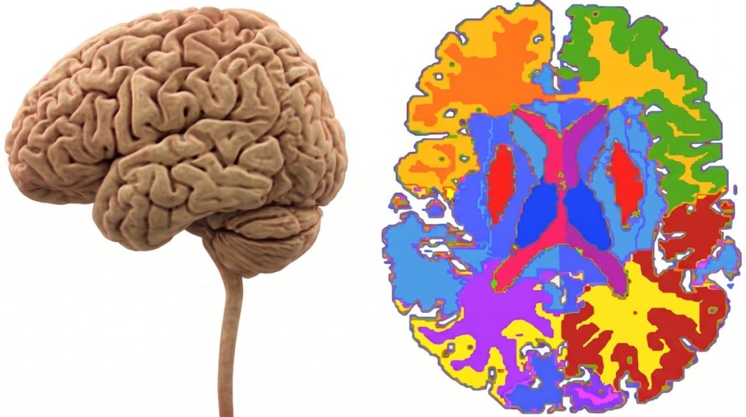
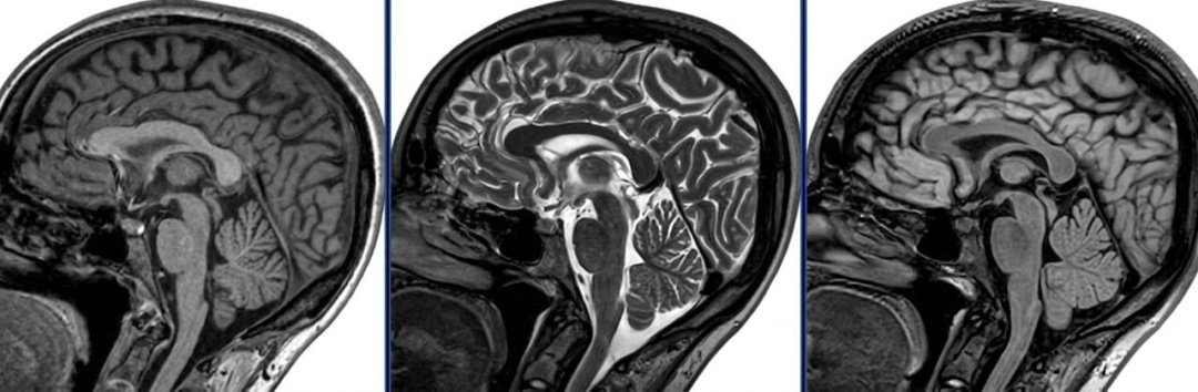
In the first stage, standard MRI scanning is used to assess the brain's overall anatomical structure and detect vascular damage. In the second stage – morphometry – a workstation automatically measures precise volumes of specific brain regions (including memory-related areas such as the hippocampus) and compares them with age-based normative databases.
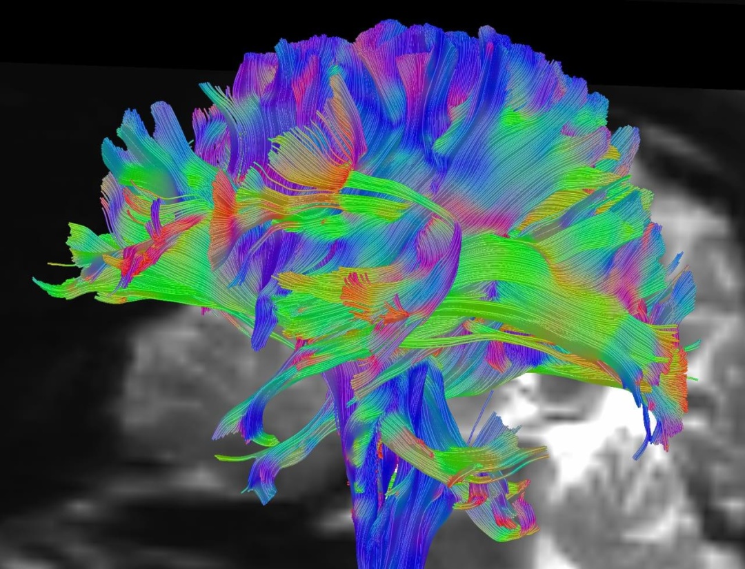
"The final stage is an analysis of the glymphatic system (DTI-ALPS), which uses mathematical algorithms to evaluate fluid movement (glymphatic clearance) along perivascular spaces, allowing the detection of stagnation processes in brain tissue," the hospital added.
The brain morphometry technology was developed by Siemens Healthineers (Germany). The concept and mathematical model for analyzing glymphatic flow were developed by a team of scientists led by Japanese professor Toshiaki Taoka in 2017.
Calculations are performed using the FSL academic software package, created by researchers at the Center for Integrative Neuroimaging at the University of Oxford (United Kingdom).
Leading medical centers worldwide (including the Mayo Clinic, Harvard Medical School hospitals, Charite, and others) use brain morphometry for the precise detection of atrophy in key brain regions, including early-stage Alzheimer's disease and other forms of dementia, while glymphatic analysis (DTI-ALPS index) is used to assess glymphatic clearance.


